What is a heart CT scan?
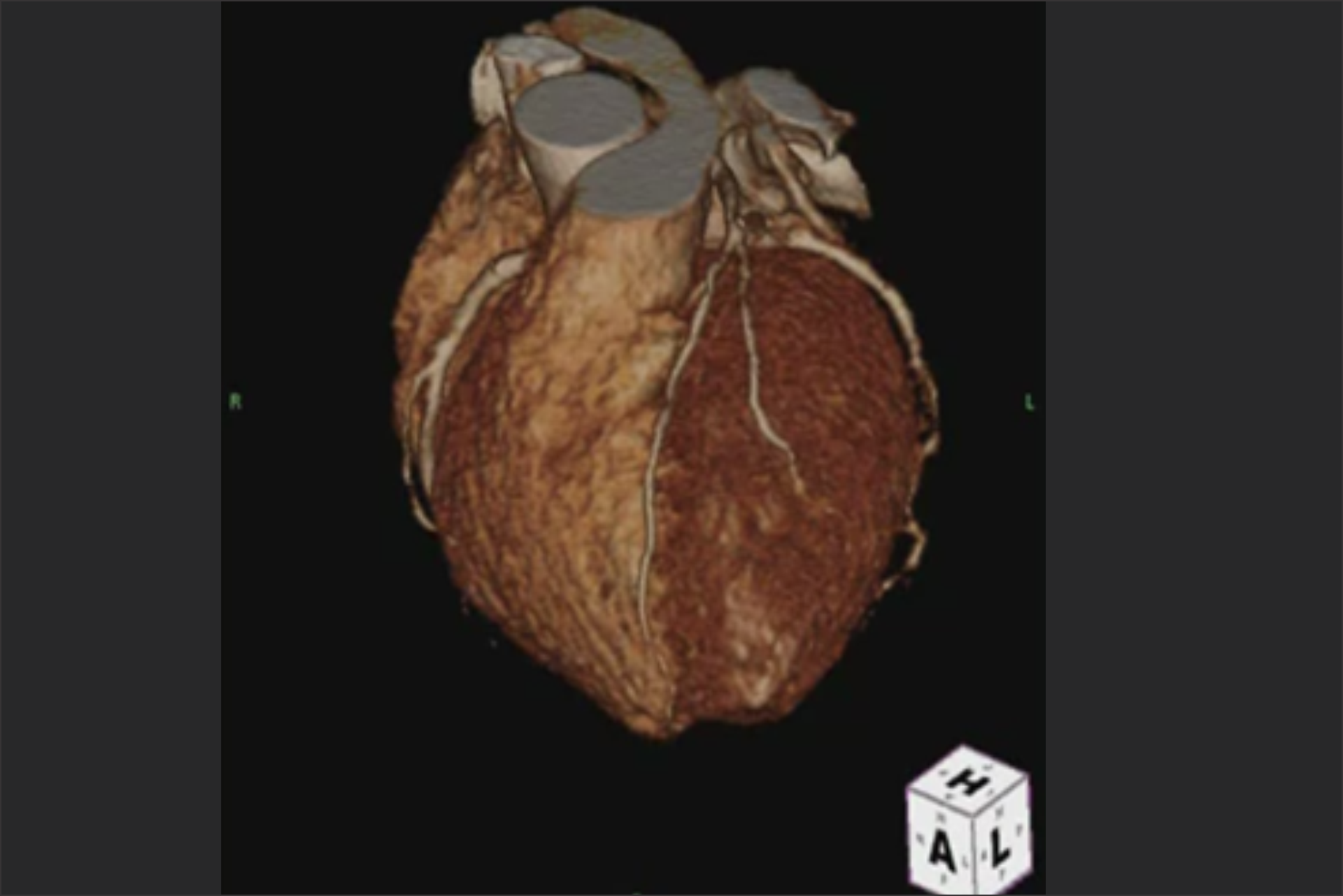
A cardiac computed tomography (CT) scan is a procedure that utilizes multiple X-ray beams from different angles to acquire high-quality, three-dimensional (3D) images of your heart, along with your great vessels and surrounding structures.
Cardiac CT uses advanced CT technology, with or without intravenous (IV) contrast (dye) to better visualize your heart structure and associated blood vessels. With multi-slice scanning, your healthcare provider can get high-resolution, 3D images of your moving heart and great vessels.
What does a CT scan of the heart show?
Your healthcare provider will be able to see your:
Coronary arteries that supply your heart.
Heart chambers, muscle and valves.
Pulmonary veins.
Thoracic aorta, and sometimes abdominal aorta.
Sac around your heart (pericardium).
When would this procedure be needed?
A cardiac CT scan can give your healthcare provider more information and detail than other kinds of imaging. Your healthcare provider may want you to have a cardiac CT scan for various reasons, including:
To evaluate the cause of chest pain and shortness of breath.
To check your heart arteries for calcium or plaque buildup, narrowing or blockages.
To assess your heart valves.
To see if there’s a problem with your aorta, including aneurysms and dissection.
To plan for open or minimally invasive/robotic heart surgery.
To plan for transcatheter/percutaneous valve procedures.
To plan for arrhythmia ablation procedures.
To assess for complications associated with the above procedures.
To see if you have a congenital (since birth) heart problem.
To see and characterize any tumor or mass in or around your heart.
To look at the sac around your heart, if there’s fluid or calcification there.